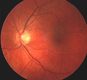
 Diabetic retinopathy is the leading cause of blindness among working ‐ age Americans. A complication of diabetes, diabetic retinopathy is an eye disease affecting the blood vessels in the retina. Long before a person notices blurring of vision from diabetic retinopathy, an eye examination can reveal abnormalities in the retina, such as the growth of abnormal blood vessels, hemorrhages (bleeding), closure of blood vessels, and leakage of fluid. In diabetics, the body can't use or store sugar properly. When blood sugar’s levels get too high, it can damage the tiny blood vessels in the retina. These small blood vessels – are especially vulnerable to the over accumulation of glucose and/or fructose that occurs in diabetes. The damage often leads to diabetic retinopathy. While not all diabetics develop Retinopathy, the longer someone has diabetes, the more likely (s) he is to have retinopathy. In fact 50% of people who have diabetes will have some vessel damage due to retinopathy within several years. In those with Diabetes for 20 years or more, that number will climb as high as 90%. In its early stages, there may be no noticeable change in vision, but left unchecked, it can lead to an advanced, sight ‐ threatening form of the disease, as damaged blood vessels in the eye become weakened, break down, or become blocked.
In many cases, diabetic retinopathy can be one of the first signs of diabetes. Anyone diagnosed with diabetes should have a dilated eye exam at least once a year.
There are two main types of diabetic retinopathy:
The early form, Non ‐ proliferative Retinopathy or background retinopathy, develops when blood vessels damaged by high blood sugar leak fluid or blood. Microaneurysms (out-growths of capillaries) occur, forming small round dark red dots on the retinal surface. As the number of microaneurysms increase, more blood vessels become blocked, and the retina is deprived of nourishment. Hemorrhages appear. The leaking fluid containing accumulations of lipids and proteins form deposits called exudates. The retina swells. If swelling occurs in the area of the macula, sight may be diminished significantly. This condition can lead to more serious forms of retinopathy that affect vision.
In Proliferative Retinopathy , fragile blood vessels develop and grow on the surface of the retina. This process is also called neovascularization. The lack of oxygen in the retina causes these fragile blood vessels to grow along the retina and in the clear, gel ‐ like vitreous ‐ humour that fills the inside of the eye. Without timely treatment, these blood vessels can bleed, cloud vision, and destroy the retina. Generally, as the vitreous becomes cloudy and prevents light from passing to the retina, the patient experiences blurry or distorted vision.
Several complications can occur as a result of diabetic retinopathy:
There are often no noticeable symptoms in the early stages of diabetic retinopathy.
The best treatment for diabetic retinopathy is prevention. People with diabetes should have their eyes checked at least annually. Good control of diabetes through the management and control of blood sugar may delay or prevent the progress of diabetic retinopathy.
Laser Photocoagulation is one of the most common treatments when diabetic retinopathy does develop. This process is highly effective in preventing visual loss from diabetic eye disease.
Focal Laser Treatment uses a laser beam to seal leaking blood vessels in the retina. This procedure may also be used to treat proliferative retinopathy by destroying abnormal blood vessels growing in the back of the eye. Patients with macular edema may be recommended to undergo focal laser photocoagulation. This entails a fluorescein angiogram to guide treatment and utilization of a laser to help “dry up” the localized swelling (macular edema). The laser treatment is applied to the macula of the eye, either in a grid‐pattern, or directly to leaking micro aneurysms. The risk of visual loss is reduced by more than 50% for patients with macular edema who undergo focal laser photocoagulation. However, vision that has been lost cannot be restored.
Scatter Laser Treatment: Rather than focus on a single spot, the laser surgeon will make hundreds of small laser burns away from the center of the retina to shrink abnormal blood vessels. Although the patient will lose some peripheral vision, the center vision will be saved. This surgery may also reduce color and night vision slightly.
Scleral Buckling (placement of an encircling band around the eye): Lasers are used to seal the detached retina to the back of the eye. A scleral buckle is a piece of silicone sponge, rubber, or semi‐hard plastic that your eye doctor sews into the scelera at the site of a retinal tear. The buckle holds the retina against the sclera until scarring seals the tear. It also prevents fluid leakage which could cause further retinal detachment. Scleral buckling is performed in an operating room under general or local anesthetic. If bleeding or inflammation blocks the surgeon's view of the retinal detachment or hole, a vitrectomy may be required.
Vitrectomy: Instead of laser surgery, some people need an eye operation called a vitrectomy to restore vision. A vitrectomy is performed when there is a lot of blood in the vitreous. It involves replacing the cloudy blood‐filled vitreous with a clear saline solution made up of salt and water which allows light to pass through to the retina. Studies show that people who have a vitrectomy soon after a large hemorrhage are more likely to protect their vision than someone who waits to have the operation. Early vitrectomy is especially effective in people with insulin‐dependent diabetes, who may be at greater risk of blindness from a hemorrhage into the eye. The procedure is done under local anesthesia as flows: After a tiny incision is made in the sclera, a small instrument is placed in the eye and is used to remove the vitreous and replace it with salt water. After surgery, the patient will need to wear an eye patch for a several days or weeks to protect the eye. Medicated eye drops are also prescribed to protect the eye.
Diabetic retinopathy cannot be prevented completely, but good control of blood sugar and healthy lifestyle can reduce the risk. Both the onset and progression of diabetic retinopathy can be slowed though control of blood sugar. Diabetics are also at higher risk for other eye diseases such as cataracts and glaucoma. A diabetic is twice as likely to get a cataract (and at an earlier age) than a non‐diabetic. They are twice as likely to develop glaucoma. Pregnant women with diabetes are at even higher risk for diabetic retinopathy. To protect their eyes, they should have eye exams every trimester.
2014. All Rights Reserved. Medical website design by Glacial Multimedia ©
The material contained on this site is for informational purposes only and is not intended to be a substitute for professional medical advice, diagnosis, or treatment. Always seek the advice of your physician or other qualified health care provider.

